
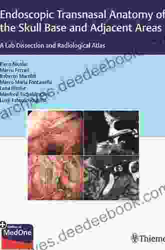
Endoscopic Transnasal Anatomy Of The Skull Base And Adjacent Areas: A Comprehensive Guide

5 out of 5
| Language | : | English |
| File size | : | 358931 KB |
| Text-to-Speech | : | Enabled |
| Screen Reader | : | Supported |
| Enhanced typesetting | : | Enabled |
| Print length | : | 784 pages |
Endoscopic transnasal surgery (ETNS) is a minimally invasive surgical technique that allows surgeons to access the skull base and adjacent areas through the nasal cavity. This approach provides direct visualization and access to anatomical structures that were previously difficult or impossible to reach using traditional open surgical techniques.
ETNS is commonly used to treat a variety of conditions affecting the skull base, including:
- Pituitary tumors
- Craniopharyngiomas
- Meningiomas
- Chordomas
- Chondrosarcomas
- Esthesioneuroblastomas
- Nasopharyngeal carcinomas
- Sinonasal tumors
In order to safely and effectively perform ETNS, surgeons must have a thorough understanding of the endoscopic transnasal anatomy of the skull base and adjacent areas.
Nasal Cavity
The nasal cavity is a three-dimensional space that is divided into two halves by the nasal septum. The roof of the nasal cavity is formed by the cribriform plate, which is a thin bony plate that separates the nasal cavity from the anterior cranial fossa. The floor of the nasal cavity is formed by the hard palate. The lateral walls of the nasal cavity are formed by the maxilla, ethmoid bone, and inferior concha.
The nasal cavity contains a number of important structures, including:
- The turbinates, which are three bony projections that project from the lateral walls of the nasal cavity and help to warm and humidify the air that we breathe.
- The olfactory bulb, which is the primary olfactory structure in the brain and is responsible for our sense of smell.
- The sphenopalatine foramen, which is a small opening in the lateral wall of the nasal cavity that transmits the maxillary nerve and vidian nerve.
- The nasolacrimal duct, which is a small channel that drains tears from the eye into the nasal cavity.
Paranasal Sinuses
The paranasal sinuses are a group of air-filled cavities that are located around the nasal cavity. The paranasal sinuses include the frontal sinus, ethmoid sinus, maxillary sinus, and sphenoid sinus.
The paranasal sinuses are lined with a mucous membrane that produces mucus. This mucus helps to keep the sinuses moist and free of infection. The paranasal sinuses also help to warm and humidify the air that we breathe.
Pituitary Gland
The pituitary gland is a small, bean-shaped gland that is located in the sella turcica, which is a bony depression in the sphenoid bone. The pituitary gland produces a number of hormones that are essential for growth, development, and metabolism.
The pituitary gland is divided into two lobes: the anterior lobe and the posterior lobe. The anterior lobe produces growth hormone, prolactin, luteinizing hormone, follicle-stimulating hormone, thyroid-stimulating hormone, and adrenocorticotropic hormone. The posterior lobe produces oxytocin and antidiuretic hormone.
Sphenoid Sinus
The sphenoid sinus is a large, air-filled cavity that is located behind the nasal cavity. The sphenoid sinus is divided into two halves by the septum of the sphenoid sinus.
The sphenoid sinus is lined with a mucous membrane that produces mucus. This mucus helps to keep the sinus moist and free of infection. The sphenoid sinus also helps to warm and humidify the air that we breathe.
The sphenoid sinus is located in close proximity to the pituitary gland, optic nerve, carotid artery, and cavernous sinus. This makes the sphenoid sinus a potential route of spread for infection and tumors.
Optic Nerve
The optic nerve is a pair of nerves that transmit visual information from the eye to the brain. The optic nerve exits the eye through the optic foramen and travels through the optic canal to reach the optic chiasm.
The optic chiasm is a small, X-shaped structure that is located at the base of the brain. The optic chiasm is where the optic nerves from each eye cross over and exchange fibers. After crossing over at the optic chiasm, the optic nerves continue on to the optic tracts.
The optic tracts are two bundles of nerve fibers that carry visual information from the optic chiasm to the visual cortex in the occipital lobe of the brain.
Carotid Artery
The carotid artery is a major artery that supplies blood to the brain. The carotid artery is divided into two branches: the internal carotid artery and the external carotid artery.
The internal carotid artery travels through the carotid canal to reach the base of the brain. The internal carotid artery supplies blood to the cerebrum, cerebellum, and brainstem.
The external carotid artery travels along the lateral wall of the neck to supply blood to the face, scalp, and neck.
Cavernous Sinus
The cavernous sinus is a large, blood-filled space that is located at the base of the brain. The cavernous sinus contains the internal carotid artery, the abducens nerve, the trochlear nerve, the oculomotor nerve, and the maxillary nerve.
The cavernous sinus is surrounded by a number of important structures, including the pituitary gland, the optic nerve, and the sphenoid sinus. This makes the cavernous sinus a potential route of spread for infection and tumors.
Clivus
The clivus is a bony ridge that is located at the base of the skull. The clivus separates the anterior cranial fossa from the middle cranial fossa.
The clivus is formed by the sphenoid bone, the occipital bone, and the petrous bone. The clivus is a potential site for chordomas and chondrosarcomas.
Petrous Bone
The petrous bone is a dense, pyramid-shaped bone that is located at the base of the skull. The petrous bone contains the inner ear and the facial nerve.
The petrous bone is a potential site for glomus tumors and paragangliomas.
Endoscopic Transnasal Surgery (ETNS)
ETNS is a minimally invasive surgical technique that allows surgeons to access the skull base and adjacent areas through the nasal cavity. ETNS is performed using a small, flexible endoscope that is inserted into the nasal cavity.
ETNS offers a number of advantages over traditional open surgical techniques, including:
- Less tissue damage
- Less pain
- Shorter recovery time
- Improved cosmesis
ETNS is commonly used to treat a variety of conditions affecting the skull base, including:
- Pituitary tumors
- Craniopharyngiomas
- Meningiomas
- Chordomas
- Chondrosarcomas
- Esthesioneuroblastomas
- Nasopharyngeal carcinomas
- Sinonasal tumors
Endoscopic transnasal anatomy is a complex and challenging subject. However, a thorough understanding of the endoscopic transnasal anatomy of the skull base and adjacent areas is essential for surgeons who perform ETNS.
By understanding the endoscopic transnasal anatomy, surgeons can safely and effectively access the skull base and adjacent areas to treat a variety of conditions.
5 out of 5
| Language | : | English |
| File size | : | 358931 KB |
| Text-to-Speech | : | Enabled |
| Screen Reader | : | Supported |
| Enhanced typesetting | : | Enabled |
| Print length | : | 784 pages |
Do you want to contribute by writing guest posts on this blog?
Please contact us and send us a resume of previous articles that you have written.
 Page
Page Reader
 Library
 Paperback
 E-book
 Magazine
 Newspaper
 Bookmark
 Shelf
Shelf Glossary
 Bibliography
 Foreword
 Synopsis
 Footnote
 Manuscript
 Scroll
 Codex
Codex Bestseller
 Classics
 Library card
 Narrative
 Biography
 Memoir
 Thesaurus
 Librarian
 Catalog
 Archives
 Study
Study Scholarly
 Lending
 Journals
 Rare Books
 Special Collections
 Interlibrary
 Study Group
 Dissertation
 Storytelling
 Reading List
 Theory
 Textbooks
 Steven P Schneider
 Graham Nuthall
 George Amberg
 Karen St James
 Philip Jacob
 Tiece
Tiece Mike Amezcua
 Frank Melling
 Mike Ormsby
 John Mccormick
 Eric C Wat
 Wade Sisson
 Caroline Braun
 Karen Baugh Menuhin
 Kenneth Kee
 Candace Fleming
 Helen Lloyd
 Erik Westhovens
 Chad Lehrmann
 Carlos Pereira Da Cruz
Light bulbAdvertise smarter! Our strategic ad space ensures maximum exposure. Reserve your spot today!


 Natsume SōsekiFollow ·12k
 Michael CrichtonFollow ·13.3k
 Maurice ParkerFollow ·12.2k
 Graham BlairFollow ·18.5k
 Francisco CoxFollow ·15k
 Italo CalvinoFollow ·2k
 Branson CarterFollow ·6.9k
 Edward ReedFollow ·10k

 Willie Blair
Lords of the White Castle: A Comprehensive Analysis of...
In the realm of...

 Edward Bell
 Dwight Bell
Fixed Effects Regression Models: Quantitative...
Fixed effects...

 Ivan Turner
Homes Around the World: A Journey Through Architectural...
Our homes are more than...

 Miguel de Cervantes
The Essentials For Standards Driven Classrooms: A...
In today's educational landscape, the...

 Colton Carter
Eugenics, Social Reform, and the Legacy of...
The early 20th century marked a period...
5 out of 5
| Language | : | English |
| File size | : | 358931 KB |
| Text-to-Speech | : | Enabled |
| Screen Reader | : | Supported |
| Enhanced typesetting | : | Enabled |
| Print length | : | 784 pages |
